
Eye Scan for Early Dementia Detection: What Corneal Confocal Microscopy Can Reveal
A painless three-minute eye scan can detect neurological disease years before symptoms appear — with higher diagnostic accuracy than an MRI. So why are neurologists still skeptical?
The window into the human nervous system has been open for 25 years. Medicine is only just beginning to look through it.
By the time a patient receives a dementia diagnosis, the neurological damage has typically been accumulating for a decade or more. The MRI scan that finally lights up reflects a brain already in crisis — a system compromised long before anyone had reason to look. Treatments initiated at that stage face steep odds. The science has known this for years. The question is why so little has been done about it.
Support Independent Local Journalism
TheTownHall.News is a non-profit reader-supported journalism. Just $5 helps us hire local reporters, investigate important issues, and hold public officials accountable across Alameda County. If you believe our community deserves strong, independent journalism, please consider donating $5 today to support our work.What Has Professor Rayaz Malik Been Building for 25 Years?
The answer, or at least a compelling part of it, sits in the research division of Weill Cornell Medicine in Qatar, where Professor Rayaz Malik has spent the better part of three decades developing a diagnostic tool that challenges the foundations of how neurological disease is detected.
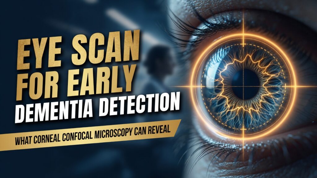
The technology is called Corneal Confocal Microscopy, or CCM. In under three minutes, a non-invasive ophthalmic scan images the nerve fibers of the cornea — the transparent front surface of the eye — at 700 times magnification. Artificial intelligence then analyzes those images to detect patterns of nerve degeneration that mirror what is happening simultaneously in the brain. Professor Malik told Dawn that researchers are using this technology to identify nerve damage years before patients begin showing symptoms. He added that AI systems can now identify the underlying neurodegenerative disease with up to 95 percent certainty.
The biological logic is precise. Corneal nerve fibers share embryological origins with the central nervous system. When neurodegeneration begins in the brain — in Alzheimer’s, Parkinson’s disease, or multiple sclerosis — it proceeds in parallel in the corneal nerves. CCM allows the corneal ultrastructure to be examined in vivo at 700× magnification, compared to the maximum 40× magnification of the standard clinical slit-lamp biomicroscope. The eye, in other words, is not a metaphor for the window into the brain. It is the actual window.
What Do the Numbers Actually Tell Us?
The scale of the problem CCM is designed to solve is staggering. There are over 55 million people worldwide living with dementia, a number set to reach 78 million by 2030 and 139 million by 2050, with the fastest growth in developing countries. In 2019, dementia cost economies globally $1.3 trillion, approximately half of that attributable to care provided by informal caregivers. In the United States alone, total payments for dementia care are projected to rise from $384 billion in 2025 to just under $1 trillion by 2050.

$1.3 trillion. The question no one in health policy wants to answer: how much of that cost traces directly to the absence of early diagnosis?
Against that backdrop, the diagnostic performance of CCM is remarkable. A study comparing CCM to MRI brain volumetry for diagnosing mild cognitive impairment and dementia found that for MCI, the area under the curve for CCM ranged from 76 to 81 percent, higher than brain volumetry at 52 to 70 percent. Translation: an inexpensive eye scan already in eye clinics worldwide outperformed the gold-standard brain imaging tool at the stage of disease when intervention could still matter most.
Research has also shown that corneal nerve loss, but not brain volumetry, was independently associated with progression from mild cognitive impairment to dementia — giving CCM higher prognostic accuracy than MRI for predicting who will deteriorate.
“The diagnostic capability of CCM is higher for identifying people with MCI and comparable for dementia — and corneal nerve loss independently predicts progression to dementia where brain MRI does not.” — Alzheimer’s & Dementia: Translational Research & Clinical Interventions [peer-reviewed data]
Is This the Accountability Moment Medicine Has Been Waiting For?
Malik published his first CCM paper in 2003. The accumulated evidence since then reaches far beyond dementia alone. CCM has been validated across Parkinson’s disease, multiple sclerosis, diabetic neuropathy, HIV neuropathy, and Friedreich’s ataxia. A study of 98 participants with Parkinson’s disease and 26 healthy controls using automated CCM analysis confirmed statistically significant reductions in corneal nerve fiber density, branch density, and fiber length in PD patients. One scan. One three-minute procedure. Multiple neurological conditions caught years earlier than any current standard allows.
If your primary care physician could detect early Alzheimer’s, Parkinson’s, and multiple sclerosis with a painless eye scan — would you want to know?
Support Independent Local Journalism
TheTownHall.News is a non-profit reader-supported journalism. Just $5 helps us hire local reporters, investigate important issues, and hold public officials accountable across Alameda County. If you believe our community deserves strong, independent journalism, please consider donating $5 today to support our work.A 2025 study published in Alzheimer’s & Dementia: Translational Research & Clinical Interventions, co-authored by Malik, confirmed that CCM detects neurodegeneration in mild cognitive impairment and dementia and identifies subjects with MCI who develop dementia. The study also investigated corneal endothelial cell morphology as an additional marker — expanding the diagnostic window further. The research continues to advance. The clinical adoption does not.
Who Is Really Blocking This Technology?
The resistance is institutional, not scientific. While CCM offers considerable promise, its widespread clinical adoption is limited by several factors, the most significant of which is the need for specialist expertise in accurate image acquisition and appropriate selection of representative frames. That barrier, however, is dissolving. AI-powered automated analysis is precisely what Malik’s team has been building — removing the need for specialist interpretation and making the scan scalable across any eye clinic with the hardware already on its shelves.
Professor Malik acknowledged that widespread adoption has faced resistance, though he noted the scan may now become widely accessible as new manufacturers begin producing the technology at lower cost.
The structural economics of medicine play a role too. MRI machines represent enormous capital investment for hospitals. Brain amyloid PET scans, now central to Alzheimer’s diagnostic protocols for new disease-modifying drugs, cost thousands of dollars per scan, are invasive by comparison, and require specialized nuclear imaging facilities. CCM requires equipment that already exists in ophthalmology clinics worldwide, costs a fraction of an MRI, and produces results in minutes. Institutional inertia rarely surrenders willingly to a cheaper, simpler competitor — even when that competitor performs better.
What Do Supporters of Current Diagnostic Protocols Actually Believe?
To be fair to the medical establishment, the skepticism around CCM is not simply protectionism or inertia. Critics raise legitimate points. Corneal nerve changes are not specific to a single disease — they occur across multiple neurological conditions, meaning a positive CCM result requires follow-up to determine which disease is progressing. Standardizing image acquisition protocols, training operators, and validating AI algorithms across diverse global populations all take time and resources. Some neurologists argue that the longitudinal evidence base — studies following patients for long enough to confirm diagnostic outcomes — is still maturing.
There is also a genuine question about what early detection means if treatment options remain limited. Traditional diagnostic approaches such as cerebrospinal fluid analysis and neuroimaging are constrained by invasiveness, high costs, and limited accessibility, particularly problematic in aging populations where early detection is crucial for effective intervention. The same argument used to defend the status quo actually underscores the case for CCM: current tools are invasive and expensive, and they still arrive too late.
The counterargument gains weight when paired with the emergence of disease-modifying therapies. FDA-approved monoclonal antibodies targeting amyloid in early Alzheimer’s require early identification of the right patients. Those drugs do not work in advanced disease. The success of such therapies relies on detecting the disease before irreversible damage in the brain occurs, requiring detection before the appearance of the first clinical symptoms. CCM, by that logic, is not just a diagnostic tool. It is the prerequisite infrastructure for an entirely new era of neurological treatment.
Are Our Leaders and Institutions Even Listening Anymore?
The fiscal argument for early detection ought to resonate in an era of government budget scrutiny. Medicare and Medicaid bear enormous dementia care costs that grow with every year of delayed diagnosis. A patient identified with mild cognitive impairment via a three-minute eye scan could be enrolled in a clinical trial, started on an emerging therapy, or placed on a structured monitoring pathway — interventions that cost far less than years of residential memory care.
The personal responsibility dimension is equally clear. Citizens who want to take charge of their own health — who believe in early action over late crisis management — are precisely the people who would benefit most from a scalable, accessible, non-invasive screening tool. The alternative is the current system: waiting for symptoms severe enough to prompt a referral, followed by a brain scan that confirms what has already been lost.
The technology exists. The evidence exists. The need exists. The only thing missing is the institutional will to act.
Professor Malik has continued presenting this evidence at major international ophthalmology conferences, most recently at the International Spectralis Symposium in May 2026, where he challenged what he called the “dogma” surrounding neurodegenerative disease diagnosis. Twenty-five years of papers, trials, and conferences. The medical establishment has had every opportunity to look through this window. The question is whether it will finally choose to.
The Real Question Is Whether Waiting Is Still Acceptable
Medicine does not fail patients only through error. It fails them through delay — through the institutional caution that holds promising tools at arm’s length while millions of families absorb the human and financial cost of late diagnosis.
Corneal Confocal Microscopy is not experimental. It is validated, peer-reviewed, and published across the world’s leading medical journals. The AI layer that enables its scalability is maturing rapidly. The equipment is already in clinics. The only missing ingredient is the decision to act — by health systems, insurers, and policymakers who still ask what the evidence says, even as the evidence stacks higher every year.
The real question is not whether this technology works. The real question is who bears responsibility for the years already lost — and whether those in a position to change the standard of care are prepared to answer for the years ahead.
Still have questions? Stay informed — subscribe for daily coverage of the health stories that matter.
Think others need to hear this? Share the article and start the conversation.
Want to make your voice count? Contact your representatives on the House Energy and Commerce Committee or Senate HELP Committee, which oversee federal health research priorities, and ask what their position is on funding early neurological screening infrastructure.
Key Questions
- Why has a technology with 25 years of peer-reviewed evidence and higher diagnostic accuracy than MRI for early cognitive impairment still not achieved widespread clinical adoption — and who is accountable for that gap?
- If FDA-approved Alzheimer’s therapies work only in early disease, and CCM can identify early disease years before MRI can, what is the actual cost — in dollars and in lives — of continuing to delay CCM integration into standard care?
- As new manufacturers lower the price of CCM equipment and AI automates its analysis, will health insurers and government programs create reimbursement pathways for early neurological screening, or will the financial incentives of the status quo hold firm?